Ventilator-associated pneumonia (VAP) is a leading cause of death in critically ill patients. Every year, many patients are diagnosed VAP. According to the NHS, 10,000 to 20,000 patients on average are diagnosed with VAP annually in the UK. Those patients with VAP have both an increased length of stay, and a higher mortality risk.
It is important for caregivers, patients and family members to understand the risks of VAP, as well as possible care techniques for reducing the likelihood of infection.
Did you know?
63%
of nurses in Europe feel they don’t have appropriate tools to provide oral care to their patients.1
VAP affects 5 to 40% of patients ventilated for more than 2 days.2
Studies suggest that VAP increases the length of ICU stay by 6 to 10 days.3
Researchers report that VAP can cost between €10,800 and €51,000 to treat.4
Main causes and risks of VAP
Main cause of VAP
When a patient has been intubated with an endotracheal or tracheostomy tube for at least 48 hours, VAP can develop. VAP is the most common ICU-acquired infection amongst mechanically ventilated patients. When patients are intubated, the coughing, chewing and swallowing actions are limited. Microorganisms can grow quickly and pool above the tracheal tube cuff. After pooling, with micro-aspiration, bacteria can enter the respiratory tract, which can develop into pneumonia.
Who is at risk?
There are certain traits, conditions or habits that may raise the risk for VAP; these conditions are known as risk factors. Risk factors for the development of VAP can be based on the patients themselves or their other diagnoses and circumstances. Risk factors can include older age, chronic lung disease, prolonged mechanical ventilation and other injuries like burns or trauma. Patients have the highest risk of developing VAP during days 1 to 5 of ventilation, and the risk is reduced each following day.5
Prevention of VAP
Elevation of the head
The position of the patient can help reduce VAP risks. If there are no other contradictions, the patient should be in semi-recumbent position (30 to 45°) as a preventive measure.6
Prevention of aspiration
VAP can be caused by aspiration of secretions that build up around the endotracheal tube in mechanically ventilated patients. The use of subglottic drainage is recommended to reduce the risk of aspiration.6
Oral care
Comprehensive oral care is vital in reducing the risk of infection. To prevent colonisation and prevent VAP, perform oral hygiene at least every 12 hours with a soft toothbrush and swabs to clean the oral mucosa and topically apply chlorhexidine gluconate (0.12% to 2%).7 Research shows that oral care protocols can reduce VAP by 46%.8
Minimisation of ventilator exposure
It is important to avoid mechanical ventilation if possible. Encouraging the use of non-invasive mechanical ventilation is one way to decrease mechanical ventilation.7 If mechanical ventilation is unavoidable, it is important to try to minimise the duration of ventilation by following evidence-based weaning protocols.7
Stress ulcer prophylaxis
Although the gastric ulcer treatment itself is not related to VAP, it does have an impact on the possible occurrence of VAP. Strong evidence suggests that lowering gastric acid for stress ulcer prophylaxis with antacids and H2 antagonists, for example, raises the risk of VAP for ventilated ICU patients.6 Where stress ulcer prophylaxis is indicated, sucralfate is recommended to reduce the risk of VAP.6
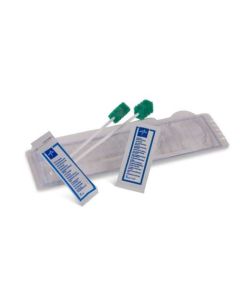
Medline VAPrevent Solutions
You can check out Medline’s oral care portfolio to help reduce the risks of VAP.

References
[1] Rello, J. et al. Oral care practices in intensive care units: A survey of 59 European ICUs. Intensive Care Medicine 33, 1066–1070 (2007).
[2] Papazian, L., Klompas, M., & Luyt, C. E. (2020). Ventilator-associated pneumonia in adults: a narrative review. Intensive Care Medicine, 46(5), 888–906. https://doi.org/10.1007/s00134-020-05980-0.
[3] Safdar N, Crnich CJ, Maki DG (2005). The pathogenesis of ventilator-associated pneumonia: its relevance to developing effective strategies for prevention. Respiratory Care 50(6):725–741.
[4] Leistner R, Kankura L, Bloch A, Sohr D, Gastmeier P, Geffers C (2013). Attributable costs of ventilator-associated lower respiratory tract infection (LRTI) acquired on intensive care units: a retrospectively matched cohort study. Antimicrobial Resistance and Infection Control 2(1):13.
[5] Pneumonia prevention systems which are designed to stop ventilator-associated pneumonia. (2022). Wessex Academic Health Science Network. https://wessexahsn.org.uk/projects/148/pneumonia-prevention-systems-which-are-designed-to-stop-ventilator-associated-pneumonia
[6] Masterton, R. G., Galloway, A., French, G., Street, M., Armstrong, J., Brown, E., Cleverley, J., Dilworth, P., Fry, C., Gascoigne, A. D., Knox, A., Nathwani, D., Spencer, R., & Wilcox, M. (2008). Guidelines for the management of hospital-acquired pneumonia in the UK: Report of the Working Party on Hospital-Acquired Pneumonia of the British Society for Antimicrobial Chemotherapy. Journal of Antimicrobial Chemotherapy, 62(1), 5–34. https://doi.org/10.1093/jac/dkn162
[7] Health Protection Surveillance Centre. (2011, February). Guidelines for the prevention of ventilator-associated pneumonia in adults in Ireland. https://www.hpsc.ie/a-z/microbiologyantimicrobialresistance/infectioncontrolandhai/guidelines/File,12530,en.pdf
[8] Sona, C. S., Zack, J. E., Schallom, M. E., McSweeney, M., McMullen, K., Thomas, J., Coopersmith, C. M., Boyle, W. A., Buchman, T. G., Mazuski, J. E., & Schuerer, D. J. E. (2008b). The Impact of a Simple, Low-cost Oral Care Protocol on Ventilator-associated Pneumonia Rates in a Surgical Intensive Care Unit. Journal of Intensive Care Medicine, 24(1), 54–62. https://doi.org/10.1177/0885066608326972